We provide the most commonly used diagnostic imaging studies in fertility, essential to evaluate the condition of the uterus, ovaries, and fallopian tubes, in order to determine the possible causes of female infertility.
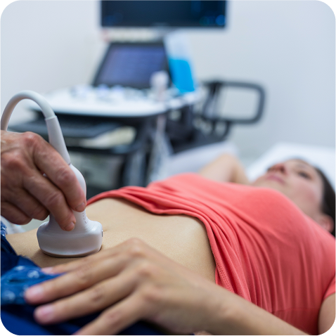
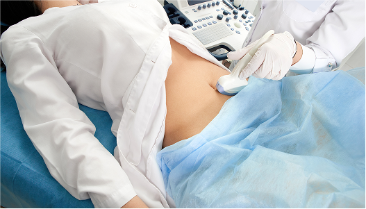
Transvaginal ultrasound (2D, 3D, sonohysterography, hysterosalpingosonography) is one of the first studies performed by the fertility specialist to evaluate the uterus, ovaries, and fallopian tubes. In addition, during treatments such as In Vitro Fertilization (IVF), several follow-up ultrasounds are performed to:
Monitor follicle development during ovarian stimulation with the help of artificial intelligence (Folliscan).
Verify the thickness and development of the endometrium before embryo transfer.
Specialized Studies in Female Fertility
We also offer other imaging studies that help provide a more detailed diagnosis:
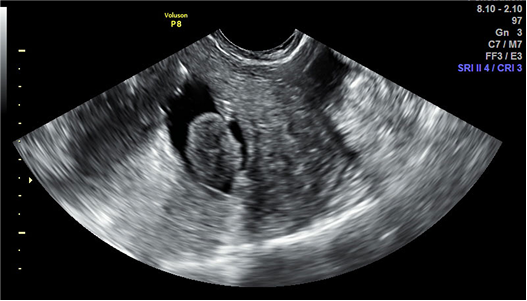
Sonohysterography:
a specialized ultrasound, where a small volume of sterile saline solution is instilled into the uttered cavity through a small catheter to detect abnormalities such as polyps, fibroids, adhesions, and deformities.
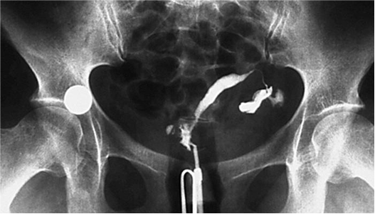
Hysterosalpingography:
an X-ray of the uterus and fallopian tubes that uses contrast media to evaluate if there are blockages or abnormalities that could hinder sperm passage or embryo implantation.